Basic techniques
| Site: | Cell Molecular Biology |
| Course: | LM-BCM Biologia Cellulare Avanzata e Biotecnologie 2015 |
| Book: | Basic techniques |
| Printed by: | Guest user |
| Date: | Friday, 7 November 2025, 12:49 AM |
1. Cell Cultures
Free online:
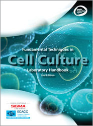
From the European Collection of Cell Culture in collaboration with Sigma Aldrich html

In the library of the Biology department:
Introduzione alle colture cellulari, 2nd edizione, Mariottini et al., Tecniche Nuove, €14,90
 previous edition:
Watch this video for basic sterile cell culture handling in sterile conditions:
Interested to watch somemore?
2. Cell count

Use of the Hemacytometer for the Determination of Cell Numbers (pdf)
Counting cells by the use of a hemacytometer is a convenient and practical method of determining cell numbers in the case that the Coulter counter is out-of-order temporarily. (It is not that bad.)
The hemacytometer consists of two chambers, each of which is divided into nine 1.0 mm squares. A cover glass is supported 0.1 mm over
these squares so that the total volume over each square is 1.0 mm x 0.1 mm or 0.1 mm3, or 10-4 cm3. Since 1 cm3 is approximately
equivalent to 1 ml, the cell concentration per ml will be the average count per square x 104.
Hemacytometer counts are subject to the following sources of error:
- Unequal cell distribution in the sample
- Improper filling of chambers (too much or too little)
- Failure to adopt a convention for counting cells in contact with the boundaries lines or with each other (be consistent)
- Statistical error
- With careful attention to detail, the overall error can be reduced to about 15%. It is assumed that the total volume in the chamber represents a random sample. This will not be a valid assumption unless the suspension consists of individual well-separated cells.
- Cell distribution in the hemacytometer chamber depends on the particle number, not particle mass. Thus, cell clumps will distribute in the same way as single cells and can distort the result. Unless 90% or more of the cells are free from contact with other cells, the count should be repeated with a new sample.
- A sample will not be representative if the cells are allowed to settle before a sample is taken. Always mix the cell suspension thoroughly before sampling.
- The cell suspension should be diluted so that each such square has between 20 - 50 cells (2-5 x 10⁵ cells/ml). A total of 300 - 400 cells should be counted, since the counting error is approximated by the square root of the total count.
- A common convention is to count cells that touch the middle lines (of the triple lines) to the left and top of the square, but do not count cells similarly located to the right and bottom.
Hemacytometer counts do not distinguish between living and dead cells.
A number of stains are useful to make this distinction. Trypan blue among others (Erythrosin B, Nigrosin) can be used: the nuclei of damaged or dead cells take up the stain. If more than 20% of the nuclei are stained, the result is probably significant. Although the trypan stain distinction has been questioned, it is simple and gives a good approximation.
Trypan blu labeling
Materials
- Clean hemacytometer and cover glass, or cover slips
- Balanced Salt Solution (BBS) or PBS
- Trypan blue, 0.4% in BBS (or PBS)
- Microscope
- Tubes
- Hand counter (Colony counter can be used)
- Cell suspension
Procedure
- Dilute 0.2 ml of Trypan blue with 0.8 ml of BBS.
- Place cover glass over hemacytometer chamber.
- Transfer 0.5 ml of agitated cell suspension to a 15 ml tube and add 0.5 ml of diluted trypan blue.
- With a 1ml pipet, fill both chambers of the hemacytometer (without overflow) by capillary action. Cells will settle in the tube and in the pipet by gravity within a few seconds. Work quickly!
- Using the microscope with a 10X ocular (and a 10X objective), count the cells in each of 10 squares (1 mm2 each). If over 10% of the cells represent clumps, repeat entire sequence. If fewer than 200 or more than 500 cells are present in the 10 squares, repeat with a more suitable dilution factor.
- Calculate the number of cells per ml, and the total number of
- cells, in the original culture as follows:
- Cells/ml = average count per square x 104
- Total cells = cells per ml X any dilution factor X total volume of cell preparation from which the sample was taken.
- Repeat count to check reproducibility (+/- 15%).
References
1. Berkson, J., T. B. Magath and M. Hurn (1939). Am. J. Physiol. 128, 309.
2. Sanford. K.K., W.R. Earle, V.J. Evans, H.K. Waltz and J.E. Shannon (1951).
3. Absher, M. in Tissue Culture Methods and Applications, Eds. Kruse, P.F. and
Patterson, M.K., Jr. Academic Press, N.Y., 1973, p.395.
From the Laboratory of Dr. Allan Bradley
Baylor College of Medicine, Houston, Texas
3. Western blotting
Theory:
SDS-PAGE
Western blotting in practice:
4. Cos cells
| Foreign Genes Can Be Expressed in Eukaryotic Cells by Utilizing Virus Transformed Cells | ||
| Figure depicts the transient expression of a transfected gene in COS cells, a commonly used system to express foreign eukaryotic genes. The COS cells are permanently cultured simian cells, transformed with an origin-defective SV40 genome. The defective viral genome has integrated into the host cell genome and constantly expresses viral proteins. Infectious viruses, which are normally lytic to infected cells, are not produced because the viral origin of replication is defective. The SV40 proteins expressed by the transformed COS cell will recognize and interact with a normal SV40 ori carried in a vector transfected into these cells. These SV40 proteins will therefore promote the repeated replication of the vector. A transfected vector containing both an SV40 ori and a gene of interest may reach a copy number in excess of 105molecules/cell. Transfected COS cells die after 34 days, possibly due to a toxic overload of the episomal vector DNA. | ||||
 | ||||
| CV1, an established tissue culture cell line of simian origin, can be infected and supports the lytic replication of the simian DNA virus, SV40. Cells are infected with an origin (ori) defective mutant of SV40 whose DNA permanently integrates into the host CV1 cell genome. The defective viral DNA continuously codes for proteins that can associate with a normal SV40 ori to regulate replication. Due to its defective ori, the integrated viral DNA will not produce viruses. The SV40 proteins synthesized in the permanently altered CV1 cell line, COS-1, can, however, induce the replication of recombinant plasmids carrying a wild-type SV40 ori to a high copy number (as high as 105 molecules per cell). The foreign protein synthesized in the transfected cells may be detected immunologically or enzymatically. | ||||